| OCR Text |
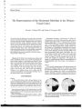
Show Journal of Neuro- Ophthalmology 20( 2): 123- 126, 2000. © 2000 Lippincott Williams & Wilkins, Inc., Philadelphia Intemuclear Ophthalmoplegia After Coronary Artery Catheterization and Percutaneous Transluminal Coronary Balloon Angioplasty Eric R. Eggenberger, DO, Nayan P. Desai, MD, David I. Kaufman, DO, and Misha Pless, MD A retrospective chart review was performed for identification of patients with isolated intemuclear ophthalmoplegia ( INO) postcardiac catheterization from two neuro- ophthalmology units. Of the 110 patients with a diagnosis of INO who were evaluated during the observation period, five patients ( 4.5%) demonstrated relatively isolated INO occurring in the perioperative period of a cardiac endovascular procedure. These five patients underwent diagnostic catheterization alone ( three patients), balloon angioplasty ( one patient), or stent placement ( one patient). All patients improved, with resolution of diplopia in primary position after a mean period of 82 days. The occurrence of INO in the postcardiac catheterization setting is not uncommon, and it appears to be related to dorsal pontine ischemia. The pontomesencephalic medial longitudinal fasciculus is supplied by small- caliber perforating end- arteries from the basilar trunk, which increases selective vulnerability of this area. Cardiac catheterization may precipitate microemboli involving these vessels, leading to intemuclear ophthalmoplegia. Key Words: Cardiac catheterization- Intemuclear ophthalmoplegia- Percutaneous transluminal coronary balloon angioplasty. Coronary artery disease ( CAD) is the leading cause of morbidity and mortality in the United States. The management of CAD has undergone a revolution in the last 20 years because of the increased use of invasive diagnostic tests and therapies. Patients with angina or suspected CAD typically undergo cardiac catheterization, endovascular therapies, including percutaneous transluminal coronary balloon angioplasty ( PTCA), and stent placement. Neurologic complications of invasive cardiac procedures include global ischemia ( hypoxic- ischemic Manuscript received January 24, 2000; accepted March 3, 2000. From the Center for Clinical Neuroscience and Ophthalmology ( ERG, NPD, DIK), Michigan State University, East Lansing, Michigan; and the Eye and Ear Institute ( MP), University of Pittsburgh Medical Center, Pittsburgh, Pennsylvania. Dr. Desai is currently affiliated with the Henry Ford Hospital Department of Neurology, Detroit, Michigan. Address correspondence and reprint requests to Eric Eggenberger, DO, Associate Professor and Co- Director, Michigan State University, Center for Clinical Neuroscience and Ophthalmology, A- 217 Clinical Center, 138 Service Road, East Lansing, MI 48824. encephalopathy), focal ischemia, and hemispheric, occipital, or brainstem infarcts ( 1,2,3). This study reports the occurrence of isolated intemuclear ophthalmoplegia ( INO) complicating endovascular cardiac procedures. METHODS The records of patients seen from 1988 through 1997 at the Michigan State University Center for Clinical Neuroscience and Ophthalmology, in East Lansing, Michigan, and the records of patients seen from 1996 through 1999 at the Eye and Ear Institute of the University of Pittsburgh Medical Center, in Pittsburgh, Pennsylvania who had a diagnosis of INO were reviewed. Inclusion criteria included the development of relatively isolated INO ( when diplopia was the primary or only symptom) related to cardiac endovascular procedures. Patients with inadequate follow- up or lacking clinical information were excluded. RESULTS Of the 110 patients with a diagnosis of INO evaluated at the two centers during the observation period, five patients ( 4.5%) fit the inclusion criteria, with relatively isolated INO occurring in the perioperative period of a cardiac endovascular procedure. These five patients underwent diagnostic catheterization alone ( three patients), balloon angioplasty ( one patient), or stent placement ( one patient) ( Table 1). No obvious patterns emerged regarding the procedure or regarding performance under emergent circumstances among the small number of patients identified. No hemorrhages were identified on imaging studies, and imaging was noncontributory in two of the four patients undergoing scans. The prognosis for recovery was generally excellent; diplopia in primary position resolved in all patients after a mean of 82 days. CASE REPORTS Case 1 A 68- year- old man experienced an acute myocardial infarction and a tissue plasminogen activator- related left 123 124 E. EGGENBERGER ET AL. TABLE 1. Clinical summary Patient/ Age/ Sex 1/ 68/ M 2/ 72/ M 3/ 53/ F 4/ 70/ F 5/ 54/ M Indication Recurrent chest pain S/ P MI Chest pain Chest pain Chest pain Chest pain Procedure/ route Cath, PTCA, stent/ femoral Cath, PTCA/ femoral Cath/ femoral Cath, PTCA/ femoral Cath/ femoral Elective/ emergent Emergent Elective Elective Emergent Elective INO: left or right Left Left Right Left Right Skew deviation left or right hypertropia Right None Left Left None Diplopia disposition 1 y ( resolved in primary gaze, 3 mo) 2 d ( resolved) 2 y ( resolved in primary gaze, 6 mo) 4 mo ( resolved) 2.5 wk ( resolved) Imaging None CT, negative; MRI, left cerebellar infarct CT, MRI, negative MRI, left pons infarct MRI, small vessel disease S/ P MI, status post myocardial infarction; Cath, catheterization; PTCA, percutaneous transluminal coronary balloon angioplasty; CT, computed tomography; MRI, magnetic resonance imaging. parietal hemorrhagic infarct with Broca aphasia. Four years later, he developed recurrent chest pain and was admitted to the hospital. An emergent cardiac catheterization was performed via the femoral approach and revealed 80% stenosis of the right coronary artery. The next day, a PTCA was performed, and stenosis was reduced to less than 20%. Within 24 hours of the procedure, the patient developed acute ST elevation, which was suggestive of an acute inferior wall infarction, and he underwent emergent stent placement in the right coronary artery. Immediately after the procedure, he noted binocular horizontal diplopia. A neuro- ophthalmic examination was consistent with a left internuclear ophthalmoplegia and skew deviation with right hypertropia. At a 3 month and at a 1- year follow- up examination, the INO pattern persisted, and the patient noted vertical diplopia only in left gaze, with no diplopia in primary position. Case 2 A 72- year- old man underwent nonemergent cardiac catheterization for evaluation of angina, and he experienced immediate postprocedure diplopia. A computed tomography ( CT) scan of brain acutely was negative. The neuro- ophthalmic examination revealed a left INO without skew deviation. This resolved in 2 days, and the patient underwent a successful and uneventful PTCA for right coronary artery stenosis. Magnetic resonance imaging ( MRI) performed 3 days after the catheterization showed a small left cerebellar infarct. At the follow- up examination 1 month later, the patient remained visually asymptomatic. Case 3 A 53- year- old woman with diabetes underwent elective cardiac catheterization for recurrent chest pains of two years' duration; the procedure was immediately followed by diplopia and left hemiparesis. Results of a CT scan and MRI were negative. A neuro- ophthalmic examination was consistent with a right INO and skew deviation ( left hypertropia). After 6 months, the diplopia resolved except in left gaze, with mild persistent left hemiparesis. At the 2- year follow- up examination, a mild right INO and skew remained without diplopia in primary position; however, diplopia persisted in left gaze. Case 4 A 70- year- old woman was admitted through the emergency department for uncontrolled hypertension and chest pain. An emergent cardiac catheterization revealed 70% stenosis of the first diagonal branch of the left anterior descending artery. A PTCA was performed 3 days later, and it was complicated by the immediate occurrence of diplopia. A neuro- ophthalmologic examination was consistent with a left INO and skew deviation ( co-mitant left hypertropia). Magnetic resonance imaging demonstrated a small region of high signal intensity in the dorsal pons within the region of the medial longitudinal fasciculus ( Fig. 1). An incidental, left sphenoid ridge meningioma was noted. At a follow- up examination 6 weeks after onset, mild left adducting lag was present; diplopia resolved 4 months after onset. Case 5 A 54- year- old man underwent elective cardiac catheterization via the femoral approach. During the proce- FIG. 1. Fluid attenuation inversion recovery MRI sequence demonstrates hyperintense lesion within the dorsal pons consistent with ischemia. J Neuro- Ophthalmol, Vol. 20, No. 2, 2000 INO AFTER CORONARY ARTERY CATHETERIZATION AND PTCA 125 dure, the patient reported diplopia. Neuro- ophthalmologic evaluation revealed a right INO without associated skew deviation or other neurologic signs or symptoms. Magnetic resonance imaging revealed small- vessel ischemic disease of the cerebral hemispheres, but results were otherwise unremarkable. The diplopia resolved within 2.5 weeks. DISCUSSION Medicine has witnessed a revolution in the treatment of coronary artery disease over the last 2 decades, with an explosion in the number of cardiac catheterizations, PTCAs, stent placements, and other invasive procedures. This trend toward more invasive testing for management of CAD will likely continue. Most of these procedures use the femoral approach, and the catheter follows a course along the arch of the aorta, passing the ostia of the left subclavian, left common carotid, and right innominate arteries as it approaches the aortic root. The occurrence of isolated INO in the postcardiac procedure setting is presumably due to ischemia referable to microembolization ( emboli < 25 u. m [ 1]), although thrombosis of the basilar perforating arteries related to preexisting vascular disease, dehydration, or hypotension may play a role. The aortic arch is a common site for friable atheroma development, especially among patients with CAD. An aortic source of cerebral emboli resulting in stroke has been elucidated using transesophageal echocardiography and spiral CT in two cases reported by Shmuely et al. ( 4). These authors described one patient with a left homonymous hemianopia and left hemipare-sis, and another patient with left hemiplegia and dysphasia. In both patients, a protruding atheroma with mobile components was demonstrated in the thoracic aorta; one was close to the origin of the left subclavian artery and another was in the ascending arch of the aorta ( 4). Although these emboli were larger and produced correspondingly larger infarctions than the relatively isolated INO observed in our patients, these data lend credence to the catheter- induced aortic emboli hypothesis. Neurologic complications after cardiac catheterization and PTCA occur at an overall rate of approximately 1.5% ( 1) and include focal ischemic neurologic deficits of anterior and posterior circulation. Single case reports of INO postcatheterization have been described among series emphasizing neurologic complications in general ( 5- 8,11,12) ( Table 2). Although the occurrence of isolated INO postcardiac catheterization appears to be quite rare, based upon existing literature, factors such as spontaneous improvement or more widespread neurologic dysfunction obscuring ocular misalignment may lead to under- recognition. Kosmorsky et al. ( 9) reported ten cases of neuro-ophthalmic complications ( two migraine and eight embolic) occurring among 30,000 cardiac catheterizations. As in other reports, the majority of the ischemic complications involved the posterior circulation,' but none of these patients had an INO. The authors speculated that the antecubital approach used in the majority of their patients played a role in the posterior circulatory preference, although clinically silent anterior circulation emboli were possible. This antecubital approach involved traversing the subclavian artery's turn at the junction of the vertebral artery. Although our patients were not studied via the antecubital approach, their deficits also involved the posterior circulation. We believe that micro-emboli are more likely to manifest clinically apparent symptoms within the posterior circulation, and the medial longitudinal fasciculus ( MLF) is a prime example of the selective vulnerability of a clinically critical structure. The prognosis for the ischemic complications in the series of Kosmorsky et al. ( 9) was generally favorable, as it was in our series. All of our patients were asymptomatic in primary position after a mean of 82 days. The MLF receives its vascular supply via the perforating arteries that originate from the posterior and lateral surfaces of the superior 1 cm of the basilar artery ( 10). These small caliber vessels demonstrate a relatively constant and predictable course traversing the brainstem in a ventral- to- dorsal manner, either through direct or circumflex branches. The direct median and paramedian vessels supply all of the midline and paramedian nuclei of the cranial nerves, including the oculomotor nuclei and the MLF. Accordingly, the MLF is supplied almost exclusively along its relatively lengthy course from the horizontal gaze center in the pons to the oculomotor nucleus in the midbrain by a series of end- arteries, where it is anatomically vulnerable to microemboli. Diplopia is a much more likely symptom to prompt neurologic or neuro- ophthalmic consultation, compared with microemboli to relatively clinically silent regions of the brain supplied by the anterior circulation. Isolated INO is a recognized complication of cardiac endovascular procedures, such as diagnostic cardiac catheterization, PTCA, or stent placement. This syndrome appears to be related to microemboli involving the unique end- artery circulation of the relatively lengthy rostral- caudal course of the MLF within the dorsal pon-tomesencephalon. Neuroimaging may be unrevealing due the limited extent of the infarction in clinically iso- Authors Dawson et al. ( 5) Galbreath et al. ( 6) Rennie et al. ( 7) Lazar et al. ( 8) TABLE 2. Year 1977 1986 1986 1995 Reports of internuclear Procedure Cath PTCA Cath Cath ophthalmoplegia Route Brachial Not stated Femoral Femoral ( INO) after cardiac procedures Duration of diplopia Not stated 7d 1 mo Not stated Cases INO/ Cases total 1/ 1,000 1/ 1,968 1 ( case report) 1/ 6,465 Cath, Catheterization; PTCA, percutaneous transluminal coronary balloon angioplasty. J Neuro- Ophthalmol, Vol. 20, No. 2, 2000 126 E. EGGENBERGER ET AL. lated cases without other neurologic signs or symptoms. The prognosis for resolution of diplopia after ischemic INO in this patient population appears to be excellent. REFERENCES 1. Furlan AJ, Sila CA, Chimowitz MI, Jones SC. Neurologic complications related to cardiac surgery. Neurol Clin 1992; 10: 145- 66. 2. Fisher- Williams M, Gottschalk PG, Browell JN. Transient cortical blindness: an unusual complication of coronary angiography. Neurology 1970; 20: 353- 5. 3. Devere TR, Lee AG, Hamill MB, et al. Acquired supranuclear oculomotor paresis following cardiovascular surgery. J Neurooph-thalmol 1997; 17: 189- 93. 4. Shmuely H, Zoldan J, Sagie A, Maimon S, Pitlik S. Acute stroke after coronary angiography associated with protruding mobile thoracic aortic atheromas. Neurology 1997; 49: 1689- 91. 5. Dawson DM, Fischer EG. Neurologic complications of cardiac catheterization. Neurology 1977; 27: 496- 7. 6. Galbreath C, Salgado ED, Furlan AJ, Hollman J. Central nervous system complications of percutaneous transluminal coronary angioplasty. Stroke 1986; 17: 616- 9, 7. Rennie IG, Wright JG, Wilkinson JL. Iatrogenic internuclear ophthalmoplegia [ letter], J Neurol Neurosurg Psychiatry 1986; 49: 842. 8. Lazar JM, Uretsky BF, Denys BG, et ai. Predisposing risk factors and natural history of acute neurologic complications of left- sided cardiac catheterization. Am J Cardiol 1995; 75: 1056- 60. 9. Kosmorsky G, Hanson MR, Tomsak RL. Neuro- ophthalmologic complications of cardiac catheterization. Neurology 1988; 38: 483- 5. 10. Hassler O. Arterial pattern of human brainstem: normal appearance and deformation in expanding supratentorial conditions. Neurology 1967; 17: 368- 75. Cited in: Hotson JR. Neurological complications of cardiac surgery. In: Miller NR, ed. Walsh and Hoyt's clinical neuro- ophthalmology. 4th ed. Vol. 4. Baltimore: Williams and Wilkins, 1991: 1912- 5. 11. Goodman GK, Crawford KH. Internuclear ophthalmoplegia following arch aortography: occurrence in association with an occult vertebral artery aneurysm. Neuroradiology 1983; 25: 171- 2. 12. Cahill DW, Salcman M, Hirsch D, Rao CV. Unilateral internuclear ophthalmoplegia due to angiographic embolism through a primitive trigeminal artery. Neurology 1981; 31: 751- 3. J Neuro- Ophthalmol, Vol. 20, No. 2, 2000 |