| OCR Text |
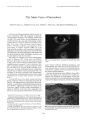
Show Journal of Neum- Ophlhulmology 18( 4): 294- 295, 1998. © 1998 Lippincott Williams & Wilkins, Philadelphia LETTERS TO THE EDITOR To the Editor: We are writing in response to the paper by Miki et al. regarding functional magnetic resonance imaging ( fMRI) of the occipital cortex in the presence of post-papilloedematous optic atrophy ( 1). The visual field illustration using automated threshold perimetry demonstrates retention of central islands of vision, left greater than right, with sparing of at least 10° of the left visual field. However, on stimulation of each eye using a flashing checkerboard at 8 Hz, no activation was produced in the striate cortex but only in the extrastriate cortex. According to the revised representation of the visual field in the occipital cortex hypothesis ( 2,3), stimulation of the left visual field should have resulted in some activation of at least 50% of the right striate cortex posteriorly. During our own studies in 12 normal healthy volunteers using fMRI we have demonstrated activation both of the striate and extrastriate cortex on stimulation of the central 11° of the visual field using a similar checkerboard at 8 Hz ( unpublished data). We therefore believe that the authors' claim that this patient's fMRI corresponded to the clinical features is not well founded. Possibly their failure to activate the striate cortex in the presence of extrastriate activation is a reflection of the use of a slice thickness of only 6 mm, given the anatomical variations of the calcarine fissure, and the small number ( only three) and duration ( not quantified) of the stimulation periods. The latter is an important parameter during activation studies, as prolonged stimulation results in an habituation- like response in the visual cortex with a decrease in the activation signal ( 4). Furthermore, the activated image in their Fig. 3 includes the superior rim of the cerebellum, which in conjunction with the roundness of the image appears to indicate that the activated slice was obtained at a steeper angle than the one usually employed to include the calcarine fissure. Unfortunately, a reference sagittal scan is not available for comparison. Thus, it may be that the activated slice does not even include the posterior striate cortex. Robert M. McFadzean, M. B., ch. B., D. O., F. R. C. Ophth. Barrie Condon, Ph. D. Institute of Neurological Sciences Southern General Hospital Glasgow, U. K. REFERENCES 1. Miki A, Nakajima T, Hasebc H, ct al. Functional magnetic resonance imaging of visual function in postpapilloedema optic atrophy. J Neuro- Ophthalmol 1997; 17: 223- 5. 2. Horton JC, Hoyt WF. The representation of the visual field in human striate cortex: a revision of the classic Holmes map. Arch Ophthalmol 1991; 109: 816- 24. 3. McFadzean R, Brosnahan D, Hadley D, et al. Representation of the visual field in the occipital striate cortex. Br J Ophthalmol 1994; 78: 185- 90. 4. Condon B, McFadzean R, Hadley D, et al. Habituation- like effects cause a significant decrease in response in MRI neuroactivation during visual stimulation. Vision Res 1997; 37: 1243^ 17. Authors' Reply To the Editor: We appreciate the comments of Drs. McFadzean and Condon. However, we have some disagreements with their interpretation of our results. In our paper, we did not state the right striate cortex of the patient had not been activated at all. The activated areas by flash stimulus are thought to be ' primitive' visual areas. Therefore, we think these areas in the occipital pole and medial occipital lobe included the striate cortex although we are not certain if they also contained V2. We selected the slice for the functional MRI after we identified the calcarine fissure in the midsagittal ' pilot' MR image. We believe our slice selection was adequate for the imaging of the calcarine cortex. In fact, we had performed functional MRI using the same slice thickness and orientation as this study in normal subjects and patients with visual deficits ( 1). In that study, we found good activation of striate cortex in normal volunteers. However, the calcarine fissure varies between individuals and sometimes winds considerably. In such cases, it may be difficult to examine the entire striate cortex by a single slice (' patchy appearance' ( 1)), and adjacent slices may also be necessary for the imaging of the striate cortex. Therefore, without a study which examines all of calcarine cortex, it is difficult to perform a ' precise' retinotopic correlation. Additionally, a rest/ stimulus cycle duration for this study is 83 seconds. This cycle duration seems to have been appropriate for the study ( 2). Atsushi Miki, M. D. Hospital of the University of Pennsylvania Philadelphia, Pennsylvania 294 |