| OCR Text |
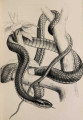
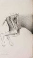
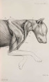
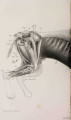
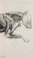
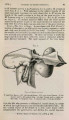
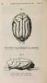
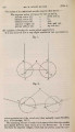
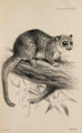
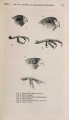
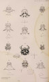
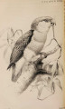
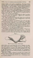
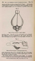
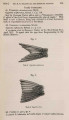
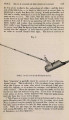
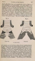
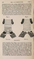
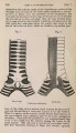
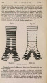
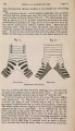
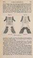
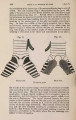
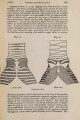
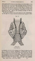
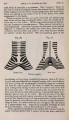
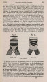
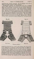
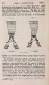
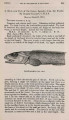
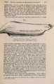
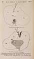
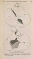
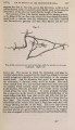
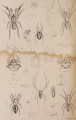
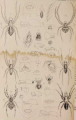
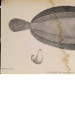
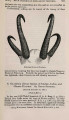
Show 1879.] ANATOMY OF HYAENA CROCUTA. 107 of place to remark that the occurrence of this divergence from the usual type, so far as its female organs are concerned, in an animal which in all other respects so closely resembles its fellows, m a y well serve to demonstrate the uncertainty of any scientific classification founded on any thing short of the consideration of the entire structure of any animal. Had the comparative anatomist examined only the female organs of H. crocuta, there can be little doubt that he would have established a separate genus, if not a family, for the reception of the animal to which they belonged. The necessity for such a course, however, is, as already pointed out, obviated by the more complete examination of the structural details of the animal. Lastly, it might be of interest to speculate as to how in the course of evolution of three species so closely allied as the three species of Hycena, two of these should have conformed to the normal mammalian type in every particular, whilst the third diverged so remarkably from that type in respect of the structural configuration of a single group of organs. Such speculations, however, do not come within the scope of a paper devoted exclusively to a record of facts. EXPLANATION OF PLATES V. & VI. Plate V. Right side of Hycena crocuta, to show tbe superficial layer of muscles drawn from the recent dissection :-G.mx., gluteus maximus, its two parts; G.md., gluteus medius; B.f, biceps femoris; T.vf., tensor vaginae femoris; Sa., sartorius, " its vertical fibres forming a superficial rectus;" P.c, panniculus carnosus; Tr., trapezius; P.,platysma ; L.d., latissimus dorsi; P.m., pectoralis major; T., triceps; L.h., levator humeri; L.s., levator scapula; D., deltoid. Plate V I . Deeper muscles, on right side of H crocuta : G.mx., gluteus maximus reflected; G.mn}, gluteus minimus, its two portions; G.mn.2, insertion of the anterior fibres of gluteus minimus; B.f, biceps femoris, reflected; G.q., gluteus quartus; Sa., sartorius; Bf., rectus femoris ; O.i., obturator interims and gemelli; Q.f, quadratus femoris; A.b., adductor brevis; A.m.+s.m., adductor magnus 4-semimembranosus; V.e., vastus externus, "booked back;" P., plantaris; Ga., gastrocnemius ; F.l.d., flexor longus digitorum ; P.I., peronaeus longus ; P.b., peronaeus brevis; E.l.d., extensor longus digitorum ; T.a.+E.l.h., tibialis anticus+extensor iongus hallucis; E.o., external oblique; B.a., rectus abdominis ; P.c, panniculus carnosus, cut; L.d., latissimus dorsi; J).e., dorsi epitrochlearis; T., triceps; T.m., teres major; S.m. ser-ratus magnus; Tr., trapezius, cut; Sc.1, Sc.2, scaleni; T.c, transversalis cervicis, " its anterior slip;" Sp., splenius; L.s., levator scapulae; L.h., levator humeri; B.a., brachialis anticus; E.c.r.b., extensor carpi radialis brevior, " and origin of longior; " E.c.d., extensor communis digitorum ; E.o.m., extensor ossis metacarpi pollicis ; E.m.d., extensor minimi digiti; E.c.u., extensor carpi ulnaris ; F.c.u., flexor carpi ulnaris; D., deltoid. |